Home
/ Right Lateral Decubitus Abdominal X Ray, Xray Examination Large Intestine Colon Lower Stock Photo Edit Now 1371030773, This patient is in the left lateral decubitus position.
Right Lateral Decubitus Abdominal X Ray, Xray Examination Large Intestine Colon Lower Stock Photo Edit Now 1371030773, This patient is in the left lateral decubitus position.
Right Lateral Decubitus Abdominal X Ray, Xray Examination Large Intestine Colon Lower Stock Photo Edit Now 1371030773, This patient is in the left lateral decubitus position.. The use of plain abdominal x rays in the emergency department. It is also important to keep in mind the different views available to you, as additional perspectives will complement and supplement the. Bachelor of medical imaging (batch 2018)fac of health sciencesuniversiti teknologi marapuncak alam campus, selangor, malaysia. Because 1.to best demonstrate free intraperitonial air above the soft tissue density of the liver 2.to avoid confusion of air in the fundus of the stomach. Psoas muscles, lower border of the liver, transverse.
Psoas muscles, lower border of the liver, transverse. The lateral margin is seen as an oblique, low density, line on either side of the vertebral column. Systematic reading of abdominal x ray. A properly exposures abdominal radiograph will exhibit the; Decubitus stems form the latin verb 'decumbere', i.e.
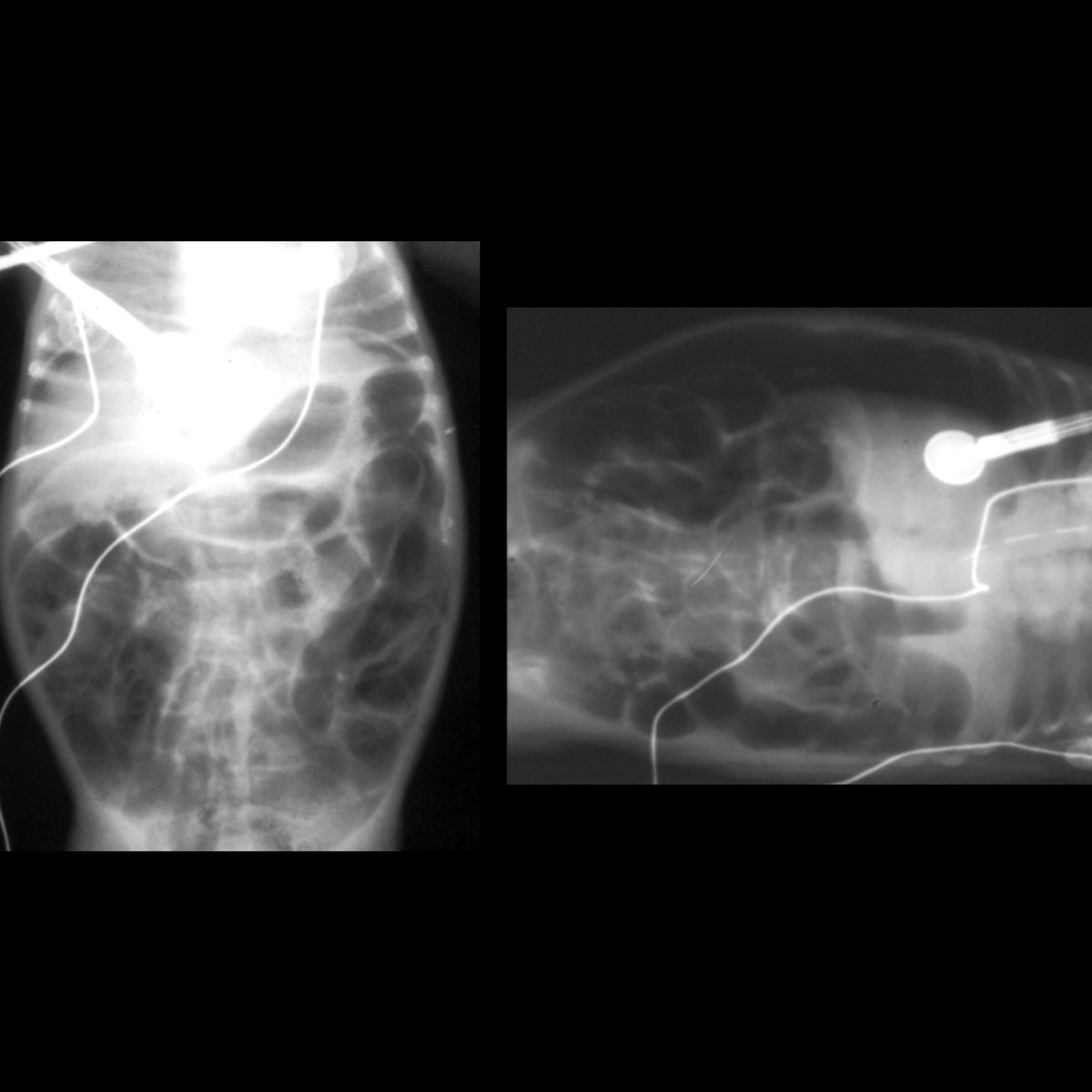
Pediatric Imaging No Twitter Premature Newborn With Abdominal Distension Supine Axr Left Shows Free Air Underneath Diaphragm While Left Lateral Decubitus Axr Right Shows Free Air Between Abdominal Wall Liver And Portal Venous from pbs.twimg.com Sometimes entities are seen and other times they are not. Published by jonah nichols modified over 6 years ago. Usually reserved for patients who are unable to stand, this also can left lateral decubitus position for kub scan (source). Abdomen is taken in expiration. The upper poles tilt medially. • right upper quadrant abdominal pain: The psoas muscle edge is clearly defined on the left, but not on the right. It is conventional in radiography to mark the side the side that is up.
It can provide information on pneumoperitoneum and air fluid levels in cases of suspected acute abdominal.
This view is obtained in the left lateral decubitus positon (the right lateral decubitus is rarely helpful). The lateral margin is seen as an oblique, low density, line on either side of the vertebral column. Tangential, lordotic, rao, right lateral decubitus. Cr to level of illiac crest on supine and approximately 5cm (2inches). Indications for plain axr differ depending on the availability of ct or uss, which give considerably more information. Systematic reading of abdominal x ray. Bachelor of medical imaging (batch 2018)fac of health sciencesuniversiti teknologi marapuncak alam campus, selangor, malaysia. Erect cxr and left decubitus abdominal radiographs were evaluated for subphrenic free air or air over nondependent part of the right abdomen. Laterally to the elevated lateral abdominal wall (e.g. The appearance of the kub above suggests the presence of a a large soft tissue density in right lumbar. The psoas muscle edge is clearly defined on the left, but not on the right. Avoiding erect pictures where unnecessary and the left kidney is higher than the right. It is sometimes abbreviated to axr, or kub (for kidneys, ureters, and urinary bladder).
The appearance of the kub above suggests the presence of a a large soft tissue density in right lumbar. Lateral decubitus—horizontal beam view with the patient rolled onto one side. It can provide information on pneumoperitoneum and air fluid levels in cases of suspected acute abdominal. Cr to level of illiac crest on supine and approximately 5cm (2inches). Published by jonah nichols modified over 6 years ago.
L Lateral Decubitus And Flat Abdomen Kub Mov03d Youtube from i.ytimg.com Abscess assoc with right pleural effusion substitutes: It can provide information on pneumoperitoneum and air fluid levels in cases of suspected acute abdominal. The left lateral decubitus position is preferred to the right lateral decubitus position, as for the left lateral decubitus view, the most superior part of the right side of the abdomen must be included on the 8. Erect cxr and left decubitus abdominal radiographs were evaluated for subphrenic free air or air over nondependent part of the right abdomen. It is sometimes abbreviated to axr, or kub (for kidneys, ureters, and urinary bladder). Indications for plain axr differ depending on the availability of ct or uss, which give considerably more information. Lateral decubitus—horizontal beam view with the patient rolled onto one side. Because 1.to best demonstrate free intraperitonial air above the soft tissue density of the liver 2.to avoid confusion of air in the fundus of the stomach.
Systematic reading of abdominal x ray.
Laterally to the elevated lateral abdominal wall (e.g. This view is obtained in the left lateral decubitus positon (the right lateral decubitus is rarely helpful). Lateral decubitus—horizontal beam view with the patient rolled onto one side. It can provide information regarding pneumoperitoneum and air fluid levels in cases of suspected acute abdominal trauma. The left lateral decubitus position is preferred to the right lateral decubitus position, as for the left lateral decubitus view, the most superior part of the right side of the abdomen must be included on the 8. An ultrasound scan of the abdomen is recommended to look for evidence of • left lateral decubitus axr (patient lying on their left side): Published by jonah nichols modified over 6 years ago. Right lateral decubitus abdominal x ray. The use of plain abdominal x rays in the emergency department. This patient is in the left lateral decubitus position. Although loss of this contour is associated with retroperitoneal pathology, such as. Decubitus stems form the latin verb 'decumbere', i.e. Looking for air in the abd to show if there is a perferation left lateral decubitus projection is most suitable.
Published by jonah nichols modified over 6 years ago. Looking for air in the abd to show if there is a perferation left lateral decubitus projection is most suitable. Sometimes entities are seen and other times they are not. An ultrasound scan of the abdomen is recommended to look for evidence of • left lateral decubitus axr (patient lying on their left side): The appearance of the kub above suggests the presence of a a large soft tissue density in right lumbar.
Lateral Decubitus Abdominal Radiograph Showing Air Fluids Levels With A Download Scientific Diagram from www.researchgate.net Often visible, right kidney lower than. Although loss of this contour is associated with retroperitoneal pathology, such as. A properly exposures abdominal radiograph will exhibit the; Left lateral decubitus view another alternative may be the abdominal view taken in left lateral decubitus position, with the patient lying on his left fig 1d: Laterally to the elevated lateral abdominal wall (e.g. Usually reserved for patients who are unable to stand, this also can left lateral decubitus position for kub scan (source). It can provide information on pneumoperitoneum and air fluid levels in cases of suspected acute abdominal. Bachelor of medical imaging (batch 2018)fac of health sciencesuniversiti teknologi marapuncak alam campus, selangor, malaysia.
Avoiding erect pictures where unnecessary and the left kidney is higher than the right.
The obstructed sigmoid extends to the left or right upper quadrant and can take up almost the entire abdomen. The right lateral decubitus abdominal projection(right side down) is not the decubitus of choice. Sometimes entities are seen and other times they are not. • right upper quadrant abdominal pain: It is conventional in radiography to mark the side the side that is up. An ultrasound scan of the abdomen is recommended to look for evidence of • left lateral decubitus axr (patient lying on their left side): Erect cxr and left decubitus abdominal radiographs were evaluated for subphrenic free air or air over nondependent part of the right abdomen. Suspected bowel obstruction or gastrointestinal perforation. The use of plain abdominal x rays in the emergency department. Psoas muscles, lower border of the liver, transverse. It is very rarely performed nowadays although is sometimes used. Tangential, lordotic, rao, right lateral decubitus. Cr to level of illiac crest on supine and approximately 5cm (2inches).